TeraRecon AV
Enterprise-level AI-enabled imaging
TeraRecon AV delivers advanced medical imaging with AI-driven automations, enhancing diagnostic precision, efficiency, and care.

Discover TeraRecon AV
AI-powered advanced visualization
Specialty care activation and coordination
Create and train your own algorithms
Advanced Visualization in practice
Step-by-step intuitive workflows
Support for device manufacturers' protocols
Increase access and availability through cloud implementation
Committed to continuous improvement
Our PACS Partners
Explore clinical solutions by body region
All Clinical Solutions
Explore every solution across each clinical category.
Cardiac CT
- Cardiac structure segmentation and functional analysis
- Automated centerline creation and vessel segmentation
- Calcium scoring with multiple database options
- Atrium and pulmonary vein analysis for EP planning
- Pre-operative evaluation of coronary arteries for plaque and stenosis
- Embedded geometry for pre-operative virtual stent evaluation
TAVR Workflow
- Aortic root segmentation and orientation
- Centerline pre-processing and extractions
- User-definable planning template
- Report output
- Semi-automated measurement protocols
LAA
- Guided LAA workflow
- Optimized cardiac orientations for the LAA
- Key measurements include landing zone, wall depth, ostium, c-arm angle, and compression measurements
- Embedded device simulation with compression percentage
- Fluoroscopy view with measurements and device
- LAA report with measurements and captured images
Cardiac MR
- Dedicated CMR Viewer
- Automatic Short Axis Contouring1
- Longitudinal Strain Analysis
- Volumetric analysis of ejection fraction
- LV/RV inner and outer contour detection
- T1, T2, T2* mapping
- AHA17-segment-model
- MR flow analysis
- Semi Quantitative MR cardiac perfusion
1Functionality requires TeraRecon Cardiac.Chambers.MR distribution
Mitral Valve/TMVR
- Guided TMVR workflow
- Optimized cardiac orientations for the mitral valve
- 2-, 3-, & 4-chamber view as well as oblique mitral valve view
- Key measurements include trigon-to-trigon distance (TT), septal-to-lateral (SL) distance, intercommissural (IC) distance measurements
- Saddle-shape or D-shape options
- Aortomitral angle measurement
Body Fusion
- Registration fusion
- Subtraction
- CT, MR, PET, SPECT
- Motion-correction
- Min, max, mean, standard deviation, standard uptake values (SUV)
- Findings viewer and follow-up
- Mirada XD Integration (Add-On)
CT Body
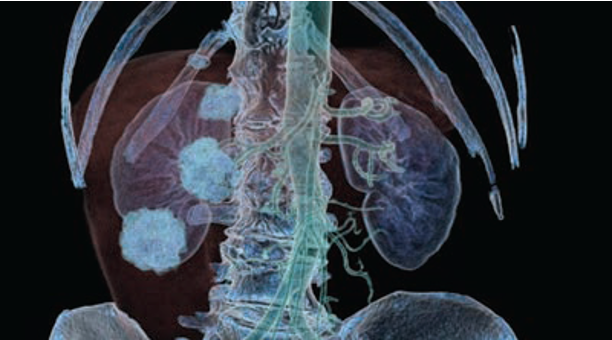
- Organ volume and histogram output
- Dynamic data support
- Sphere-like structure identification
- Dynamic image filtering
- Dual-source data support
- Colon flythrough
- Streamlined screening
- Enhanced clinical confidence
- Personalized care plans
MR Body
- 2D, 3D, 4D MR image sequencing
- MIP and MRA evaluation with centerline tools
- Analysis and follow-up tools
- Time-intensity ROI analysis
- Parametric mapping of body parts such as breast or prostate
- Kinetics, time-to-peak, time to the enhancement and maximum slope evaluation
- Streamlined screening
- Enhanced clinical confidence
- Personalized care plans
Lung Segmentation
Lung and trachea segmentation
Lung and lobe volumetric analysis
Lung, airway, and vessel anatomical fusion
Treatment planning simulation
Low attenuation
Lung Density Analysis II
Semo-automated lung segmentation
Lung volume and histogram analysis
Sphere-like structure identification
Comparative tracking-over-time options
Virtual flythrough
Rib Labeling
CT Chest
Semi-automated lung segmentation
Lung volume and histogram analysis
Sphere-like structure identification
Comparative tracking-over-time options
Virtual flythrough
Rib Labeling
Maxillofacial
Panoramic projection
Cross-sectional multi-planar reconstruction
Definable mandibular groove path
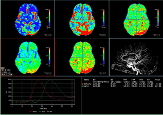
Neuro CT Perfusion Workflow*
Tmax
Mismatch
Hypoperfusion Maps
*Results provided from TeraRecon Neuro
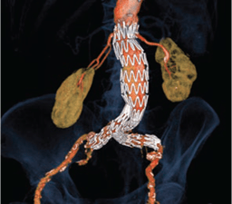
EVAR Workflow

- Pre-generated centerlines
- User-definable planning template
- Diameter vs. distance and cross-sectional views
- Straightened view, diameter, and length measurements
- Embedded vendor-specific report templates
- Streamlined screening
- Enhanced clinical confidence
- Personalized care plans
Interventional Radiology

Centerline analysis tools
Stent-graft planning
Curved planar reformation
Analysis and follow-up tools
Perspective flythrough
- Streamlined screening
- Enhanced clinical confidence
- Personalized care plans
CT Head and Neck

- Automatic centerline identification simplifies stenosis measurements
- Dual source data support
- Time density evaluation: maps include CBF, CBV, MTT, TTP, Tmax*, hypoperfusion*, mismatch*, and more
- Multi-modality image fusion and image subtraction
*Results provided by TeraRecon Neuro
- Streamlined screening
- Enhanced clinical confidence
- Personalized care plans
Alignment Measurements
- TT-TG measurement
- Cobb angle measurement
Vertebral Labeling Workflow
- One click center line creation
- True axial
- Coronal reformats
Rib Labeling Workflow
- Rib Labeling
- Centerline generation
- Fishbone image display
- Efficient fracture detection and evaluation
Cardiology
Tools for cardiovascular imaging workflows.
Cardiac CT
- Cardiac structure segmentation and functional analysis
- Automated centerline creation and vessel segmentation
- Calcium scoring with multiple database options
- Atrium and pulmonary vein analysis for EP planning
- Pre-operative evaluation of coronary arteries for plaque and stenosis
- Embedded geometry for pre-operative virtual stent evaluation
TAVR Workflow
- Aortic root segmentation and orientation
- Centerline pre-processing and extractions
- User-definable planning template
- Report output
- Semi-automated measurement protocols
LAA
- Guided LAA workflow
- Optimized cardiac orientations for the LAA
- Key measurements include landing zone, wall depth, ostium, c-arm angle, and compression measurements
- Embedded device simulation with compression percentage
- Fluoroscopy view with measurements and device
- LAA report with measurements and captured images
Cardiac MR
- Dedicated CMR Viewer
- Automatic Short Axis Contouring1
- Longitudinal Strain Analysis
- Volumetric analysis of ejection fraction
- LV/RV inner and outer contour detection
- T1, T2, T2* mapping
- AHA17-segment-model
- MR flow analysis
- Semi Quantitative MR cardiac perfusion
1Functionality requires TeraRecon Cardiac.Chambers.MR distribution
Mitral Valve/TMVR
- Guided TMVR workflow
- Optimized cardiac orientations for the mitral valve
- 2-, 3-, & 4-chamber view as well as oblique mitral valve view
- Key measurements include trigon-to-trigon distance (TT), septal-to-lateral (SL) distance, intercommissural (IC) distance measurements
- Saddle-shape or D-shape options
- Aortomitral angle measurement
Body imaging/oncology
Precise imaging workflows to aid complex cancer treatments.
Body Fusion
- Registration fusion
- Subtraction
- CT, MR, PET, SPECT
- Motion-correction
- Min, max, mean, standard deviation, standard uptake values (SUV)
- Findings viewer and follow-up
- Mirada XD Integration (Add-On)
CT Body
- Organ volume and histogram output
- Dynamic data support
- Sphere-like structure identification
- Dynamic image filtering
- Dual-source data support
- Colon flythrough
- Streamlined screening
- Enhanced clinical confidence
- Personalized care plans
MR Body
- 2D, 3D, 4D MR image sequencing
- MIP and MRA evaluation with centerline tools
- Analysis and follow-up tools
- Time-intensity ROI analysis
- Parametric mapping of body parts such as breast or prostate
- Kinetics, time-to-peak, time to the enhancement and maximum slope evaluation
- Streamlined screening
- Enhanced clinical confidence
- Personalized care plans
Pulmonology
Reliable Tools to Aid in Addressing Pulmonary Disease
- CT Chest
- Lung Segmentation
- Lung Density Analysis II
- Diagnostic support
- Streamlined workflows
- Precise planning for interventions
- Faster decision making & better patient management
Lung Segmentation
Lung and trachea segmentation
Lung and lobe volumetric analysis
Lung, airway, and vessel anatomical fusion
Treatment planning simulation
Low attenuation
Lung Density Analysis II
Semo-automated lung segmentation
Lung volume and histogram analysis
Sphere-like structure identification
Comparative tracking-over-time options
Virtual flythrough
Rib Labeling
CT Chest
Semi-automated lung segmentation
Lung volume and histogram analysis
Sphere-like structure identification
Comparative tracking-over-time options
Virtual flythrough
Rib Labeling
Neurology
Stroke Care, Timely Automation, and Optimized Neuro Disorder Workflow
- CT Head & Neck
- Brain Perfusion
- Maxillofacial
Customer Benefits
Faster patient assessment
Improved collaboration between neurology and radiology
Stroke intervention insights
Single, cohesive AI-enabled AV solutions
Maxillofacial
Panoramic projection
Cross-sectional multi-planar reconstruction
Definable mandibular groove path
Neuro CT Perfusion Workflow*
Tmax
Mismatch
Hypoperfusion Maps
*Results provided from TeraRecon Neuro
Vascular
Informed Diagnosis & Planning for Complex Situations
EVAR
Interventional Radiology
Calcium Workflow
Aorta.CT, AI-automated Workflow*
*Requires TeraRecon Aorta.CT
Customer Benefits
Automated segmentation & labeling
Enhanced planning for interventional procedures
Single, cohesive AI-enabled AV solutions
EVAR Workflow

- Pre-generated centerlines
- User-definable planning template
- Diameter vs. distance and cross-sectional views
- Straightened view, diameter, and length measurements
- Embedded vendor-specific report templates
- Streamlined screening
- Enhanced clinical confidence
- Personalized care plans
Interventional Radiology

Centerline analysis tools
Stent-graft planning
Curved planar reformation
Analysis and follow-up tools
Perspective flythrough
- Streamlined screening
- Enhanced clinical confidence
- Personalized care plans
CT Head and Neck

- Automatic centerline identification simplifies stenosis measurements
- Dual source data support
- Time density evaluation: maps include CBF, CBV, MTT, TTP, Tmax*, hypoperfusion*, mismatch*, and more
- Multi-modality image fusion and image subtraction
*Results provided by TeraRecon Neuro
- Streamlined screening
- Enhanced clinical confidence
- Personalized care plans
MSK
Precise Imaging Workflows to Aid Complex Treatments
Vertebral Labeling Workflow
Rib Labeling Workflow
Alignment Measurements
Customer Benefits
Semi-automated vertebral and rib labeling across axial, sagittal, coronal, and 3D views dramatically reduces manual effort while accelerating interpretation workflows.
Dynamic, intuitive visualization tools bring complex anatomy to life—making joint disarticulation fast, precise, and effortless.
Advanced 2D and 3D anatomical visualization and measurement tools empower confident decision-making for trauma fixation, angle and length assessment, bone alignment, joint space evaluation, Cobb angles, TT-TG, and much more.
Alignment Measurements
- TT-TG measurement
- Cobb angle measurement
Vertebral Labeling Workflow
- One click center line creation
- True axial
- Coronal reformats
Rib Labeling Workflow
- Rib Labeling
- Centerline generation
- Fishbone image display
- Efficient fracture detection and evaluation