Customer Services
Learning Center
About Us
Customer Services
Learning Center


.svg)

Over the last few months, stories about Traumatic Brain Injury (TBI) have been very popular across the full spectrum of news media. There are articles in newspapers, national news magazines and segments on local & national news. These stories have wide appeal because this subject affects so many of us. People injured in automobile accidents or falls, veterans returning from service and children playing school sports may all be at risk.
 So what exactly is TBI? Traumatic Brain Injury can most readily be defined as a physical injury to the brain. It usually occurs when the brain encounters force, typically via direct impact or indirect impact. Examples of indirect impact are whiplash or the concussive blast from an explosion. In both of these examples force is applied to the brain even though no direct contact between the head and a physical object occurs. Many of the events we see discussed in the media as potential causes of TBI are commonly referred to as concussions.
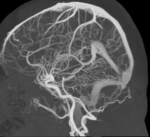
TBI is not a disease, it is a clinical diagnosis that must be determined by a physician. Current practice is to apply the Glasgow coma scale. This approach is largely symptom-based and most frequently confirmed with correlated findings from CT or MRI exams. Mild cases of TBI can be difficult to diagnosis as CT and MRI of the head exams frequently come back negative in instances of mild TBI. As an aid to the Neuro-Radiologist in making this difficult diagnosis, TeraRecon’s Intuition with TDA (Time Dependent Analysis) provides the ability to analyze dynamic data to support the assessment of time-dependent behavior of the image intensity or density of the brain including CBF, CBV, MTT, TTP, TOT, RT and uptake graph (Time Density Graph).  Intuition supports data from dual source CT scanning and can provide dynamic blending. Intuition also provides tools to reduce image noise and subtraction of high density structures, such as bone and metal.
In mild TBI, a definitive image-based finding can be very elusive. A promising new technique has recently been suggested by Dr.Gerald Riedy from Walter Reed. He suggests using a 3T MR to acquire T1/T2 3D volume imaging at sub 1mm slices to produce highly detailed images of the brain which may reveal small white matter T2 weighted hyper-intensities that may be clinically significant, especially when compared to the age of the patient. 2 Dr.Riedy also recommends using GRE susceptibility imaging, which may reveal ‘blooms’ indicative of micro-hemorrhage. There is also a great deal of promise reported in Dr.Riedy’s findings related to fMRI in diagnosis of TBI. Hopefully, his innovative research will lead the way to new imaging techniques that will enable the Radiologist to more readily identify this often elusive diagnosis.
Please click here to learn more about Dr.Riedy’s exciting new imaging techniques.
1 http://www.mayoclinic.org/diseases-conditions/traumatic-brain-injury/basics/symptoms/con-20029302
2 http://www.rsna.org/News.aspx?id=18115
https://www.ncbi.nlm.nih.gov/pmc/articles/PMC3586697/
http://www.today.com/video/is-football-safe-for-kids-study-looks-at-brain-changes-792170051980
Leading Causes of TBI Image Source: https://www.cdc.gov/traumaticbraininjury/get_the_facts.html

No Comments Yet
Let us know what you think